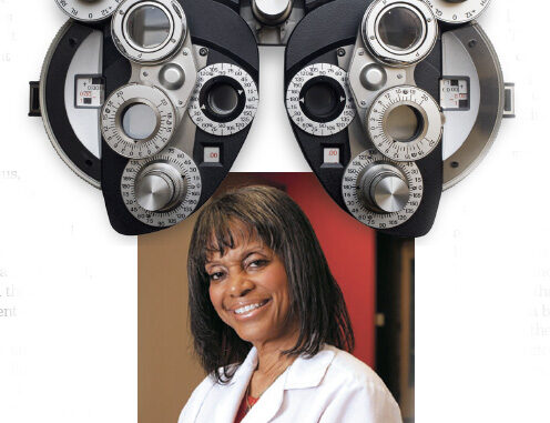
By Dr. Paula Newsome
There are a few ocular emergencies that you could experience and retinal detachment is one of them.
The retina is the thin lining of the inside of the eye and is the only place in the body where we can directly look at arteries, veins and nerves without physically cutting. It also is the home of the light sensing and the seeing part of our visual system. While it is paper thin in appearance it contains 10 layers which are critically important in seeing and thus, make retinal detachment a true ocular emergency.
The retina is part of the brain and central nervous system and like the spinal cord, once the retina is damaged, it does not regenerate and thus a complete retinal detachment can cause permanent blindness.
So, what are some signs and symptoms of a retinal detachment? Flashes of light that appear like lightning flashes. Another sign is seeing floaters which can appear like little flecks through your field of vision. Blurry vision can also be a sign of a retinal detachment especially if it’s a sudden loss of vision or sudden blurring of vision. Gradually reduced side vision or peripheral vision may also be a sign of a retinal detachment. Also, a curtain or a veil-like shadow over your vision may also be a sign of a detachment. Any of these signs are a cause for concern and warrant an in-person visit to your eye doctor for a dilated examination.
There are three main types of retinal detachments. The more common one is the rhegmatogenous detachment. This type of detachment is where a hole or tear in the retina allows fluid to seep behind it and thus causes the retina to separate from its normal position. If caught early and depending on where the hole is, these detachments can be repaired. If, however, they go undetected or undiagnosed, a complete and total detachment can occur and this could lead to permanent vision loss.
Tractional detachment is most frequently found in people with uncontrolled diabetes. It is where tissue grows on the retinal surface and that tugs the retina away. This is yet another reason why people with diabetes should have their eyes checked on a regular basis.
The last type of detachment occurs because fluid caused by disease, tumors or inflammatory disorders get between the retina and the underlying vascular layer of the eye, the choroid. This last type of detachment is called exudative and is seen in people with tumors, macular degeneration and some other inflammatory diseases.
In addition to signs and symptoms, there are also some risk factors for getting retinal detachments. Trauma is one that immediately comes to mind, including any type of physical insult to the eye or head. Aging is another risk factor. As we have more and more birthdays, the gel in our eyes that is immediately in front of the retina becomes liquid and that can tug on the retina, pulling it away. Previous history of a retinal detachment makes you more susceptible to having a detachment in the other eye. Family history of a retinal detachment also is a risk factor. If people in your family have had a retinal detachment, you need to let your eye care provider know. Any eye surgery or any eye diseases can make you more susceptible. High myopia or high nearsightedness can also make you more susceptible to a retinal detachment because, often times, the retina must stretch around a larger area.
There are no home remedies for this problem. The most important preventative tip is to make sure that you have your eyes examined regularly. During the examination, you should either be dilated or have wide-field photography to make sure there are no holes or tears in your eye’s periphery.
You only get two eyes, make sure you take care of them for a lifetime of good vision.
Dr. Paula Newsome is the first African American woman optometrist to open a private practice in North Carolina and owner of Advantage Vision Center, 1601 S. Church St., Charlotte, N.C.
